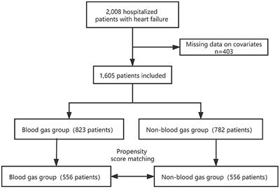
Patient flow diagram. A total of 556 patients who were classified as

Download scientific diagram | Patient flow diagram. A total of 556 patients who were classified as either stage C or D of HF stage classification in the ACCF/AHA guidelines and were hospitalized at Fukushima Medical University Hospital between April 2018 and September 2021 underwent abdominal ultrasonography. Patients who had chronic liver disease (eg, viral hepatitis, cirrhosis, hepatic tumors, bile duct disease, alcohol abuse) or were undergoing dialysis were excluded from the study. Finally, 513 patients were enrolled, among whom 310 had undergone right heart catheterization. Of these patients, those with HVSI of 0 were classified as the HVSI 0 group. Patients with HVSI above 0 were divided into 2 groups based on the median HVSI value (0.20) in the present population: the low HVSI group and the high HVSI group. ACCF indicates American College of Cardiology Foundation; AHA, American Heart Association; HF, heart failure; and HVSI, hepatic venous stasis index. from publication: Hepatic Venous Stasis Index Reflects Hepatic Congestion and Predicts Adverse Outcomes in Patients With Heart Failure | Background It has been reported that the hepatic vein waveforms determined by abdominal ultrasonography can assess hepatic congestion in patients with heart failure (HF). However, the parameter that quantifies hepatic vein waveforms has not been established. We suggest the | Congestion, Stasi and Heart Failure | ResearchGate, the professional network for scientists.

Discharge of postoperative patients with an opioid prescription is
Frontiers The Relationship Between the Utilization of Arterial

Patient Flow Chart – Insights, Templates, and Creation Procedure
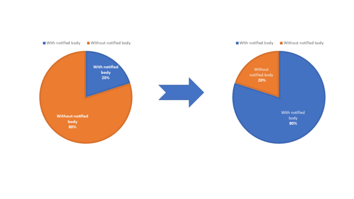
Classification of in-vitro diagnostic medical devices

Masayoshi Oikawa's research works Fukushima Medical University, Fukushima and other places

Patient Flow Chart – Insights, Templates, and Creation Procedure

Non-Small Cell Lung Cancer Treatment (PDQ®) - NCI
Patient identification flowchart. ICD-9, International

Patient inclusion. The figure illustrates a flowchart of patient

Risk factors for death caused by early onset sepsis in neonates: a
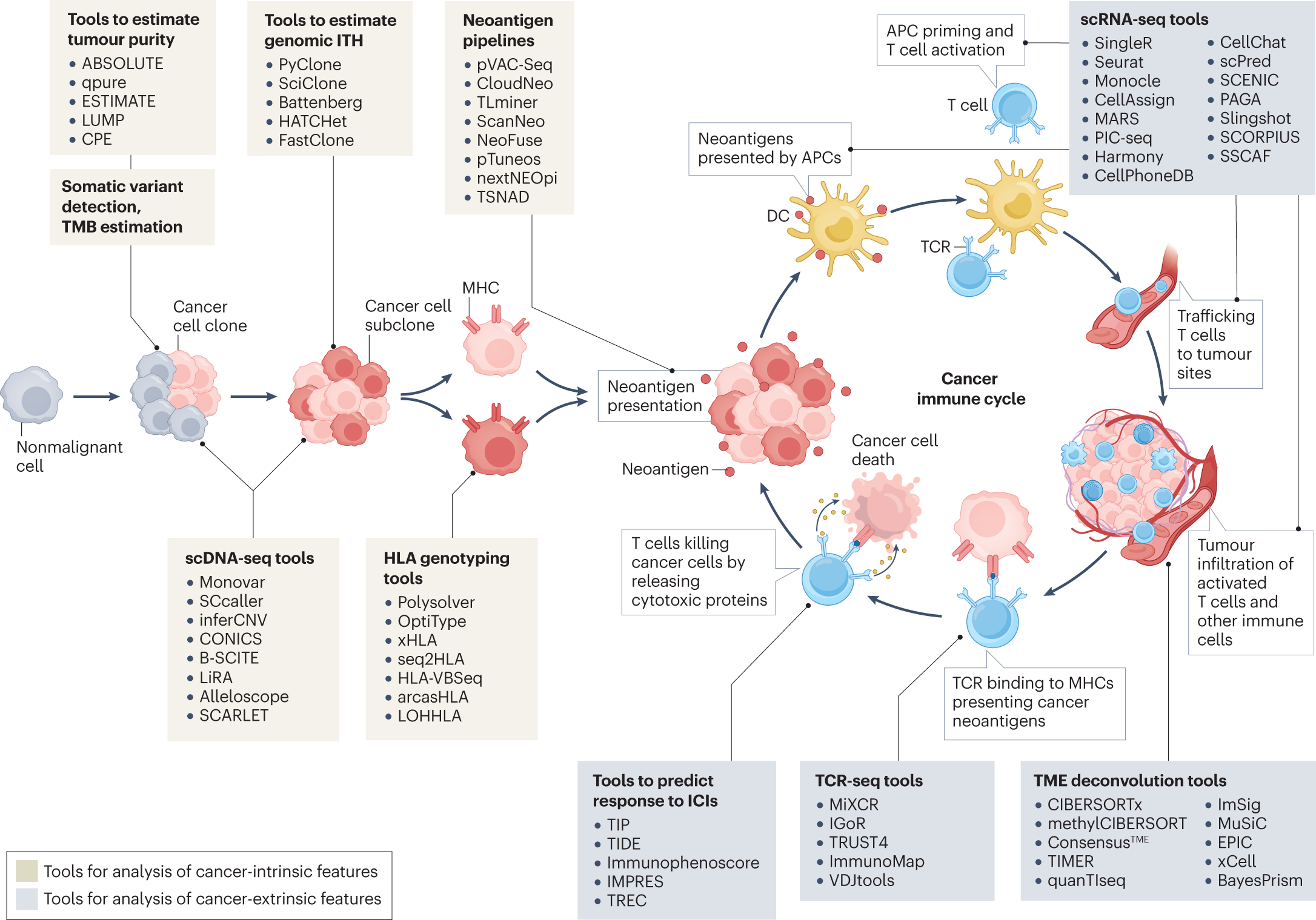
Computational immunogenomic approaches to predict response to
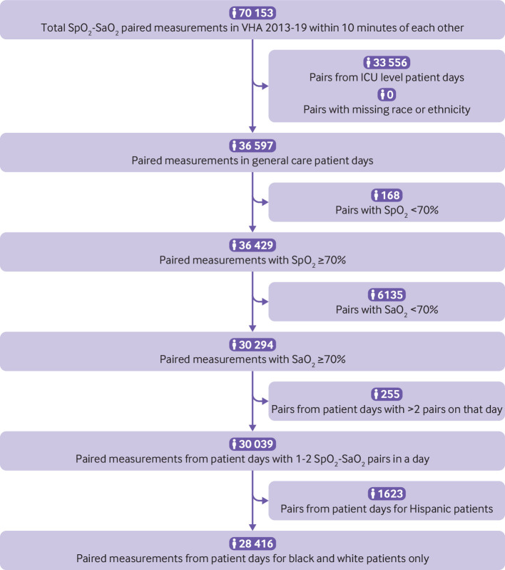
VHA Pulse Oximeters Over-Estimated Oxygenation in Some Racial
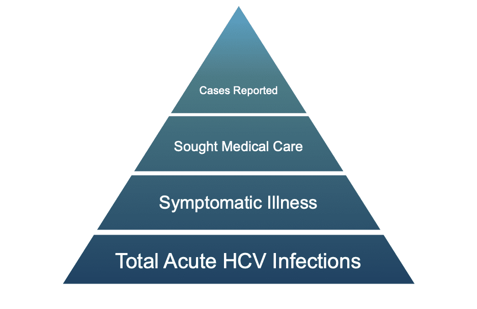
Core Concepts - HCV Epidemiology in the United States - Screening
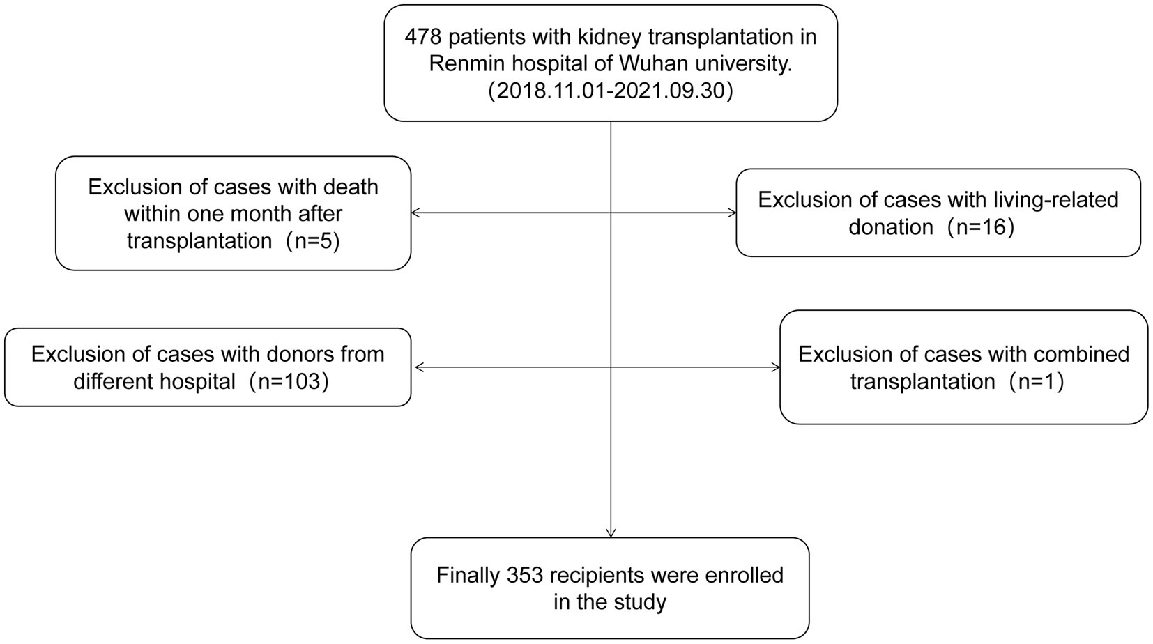
Frontiers Risk factors for BK virus infection in DCD donor