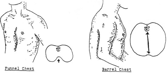
Bell-shaped chest is demonstrated with small upper chest


Venkatraman BHAT, Senior Consultant, Imaging

PDF) Further Observations in Cerebro-Costo-Mandibular Syndrome: Exploration with CT Imaging

Bell-shaped chest is demonstrated with small upper chest. Musculature

Venkatraman BHAT, Senior Consultant, Imaging

Venkatraman BHAT, Senior Consultant, Imaging

Venkatraman BHAT, Senior Consultant, Imaging

Venkatraman BHAT, Senior Consultant, Imaging

PDF) Further Observations in Cerebro-Costo-Mandibular Syndrome: Exploration with CT Imaging

Bell-shaped chest is demonstrated with small upper chest. Musculature

PDF) Further Observations in Cerebro-Costo-Mandibular Syndrome: Exploration with CT Imaging

PDF) Further Observations in Cerebro-Costo-Mandibular Syndrome: Exploration with CT Imaging

PDF) Further Observations in Cerebro-Costo-Mandibular Syndrome: Exploration with CT Imaging
Bell-shaped chest is demonstrated with small upper chest. Musculature


:max_bytes(150000):strip_icc()/iStock_20479601_MEDIUM-582cd9df3df78c6f6aaaae0c.jpg)